104
The Morphology of Human Blood Cells
Dorothy Sturm
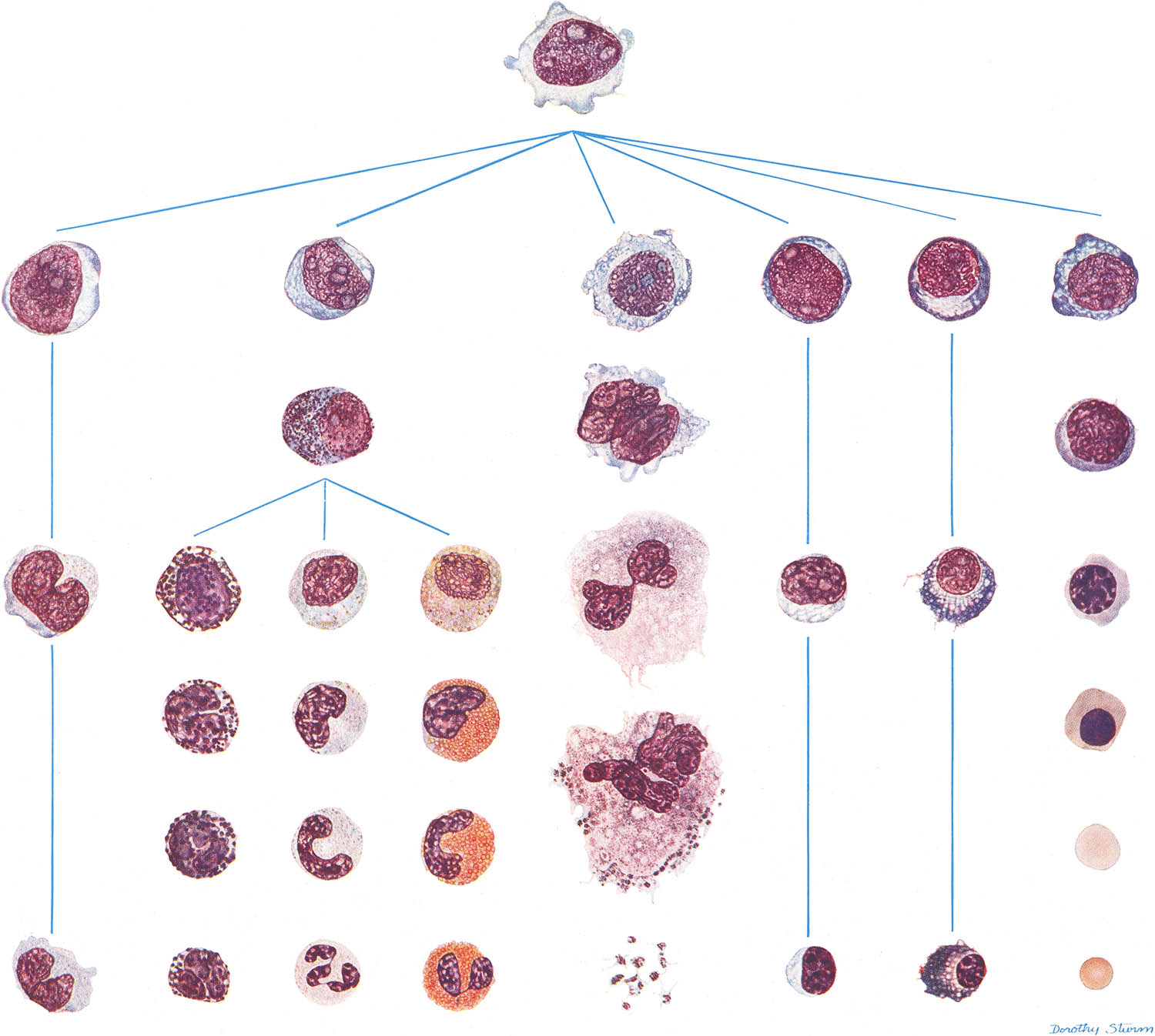
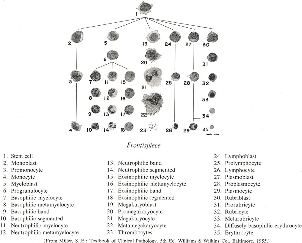
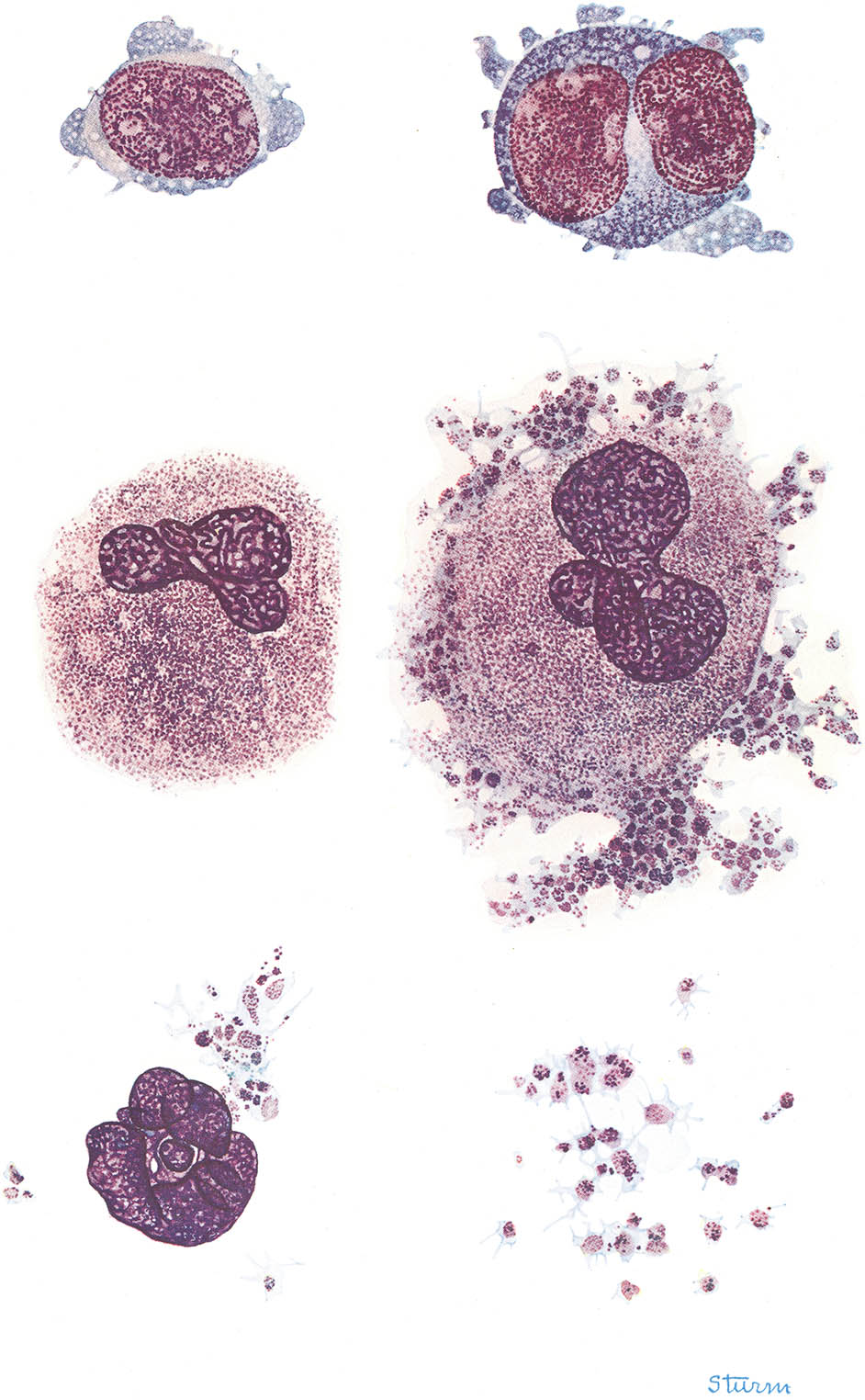
This table showing hematopoiesis (as it was understood in 1956) was the frontispiece of the first edition of Diggs’ The Morphology of Human Blood Cells. At first glance Dorothy Sturm’s beautiful watercolors are rather difficult to tell apart from an actual microphotograph (except perhaps they are clearer and more detailed). For those playing along at home here is the key:1
Dorothy Sturm was born in 1910 in Memphis.2 At 19 she moved to New York and studied at the Grand Central School of Art and the Art Students League. She became interested in the work of the pioneering Dr. Florence Sabin and with the addition of biology studies at Columbia became a free-lance medical illustrator.
She returned to Memphis in 1924 as an instructor at the Memphis Academy of Arts. Sturm, described in a 1936 UPI article as “attractive, with black curly hair and blue eyes,”3 was soon in high demand by regional physicians and surgeons to illustrate books, articles and papers.
It’s no surprise then that when the University of Tennessee hematologist Lemuel Diggs4 and his assistant Ann Bell began working on their atlas of hematology – The Morphology of Human Blood Cells5 – they commissioned Sturm to provide the illustrations.
Maturation sequence
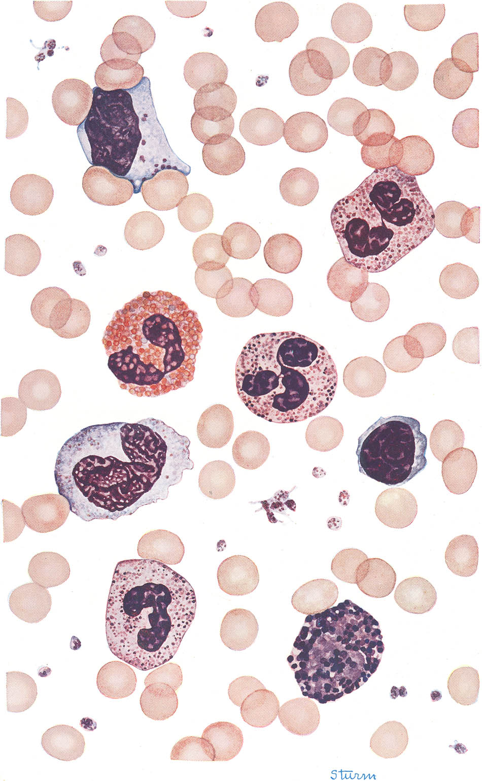
Cell types found in smears of peripheral blood from normal individuals
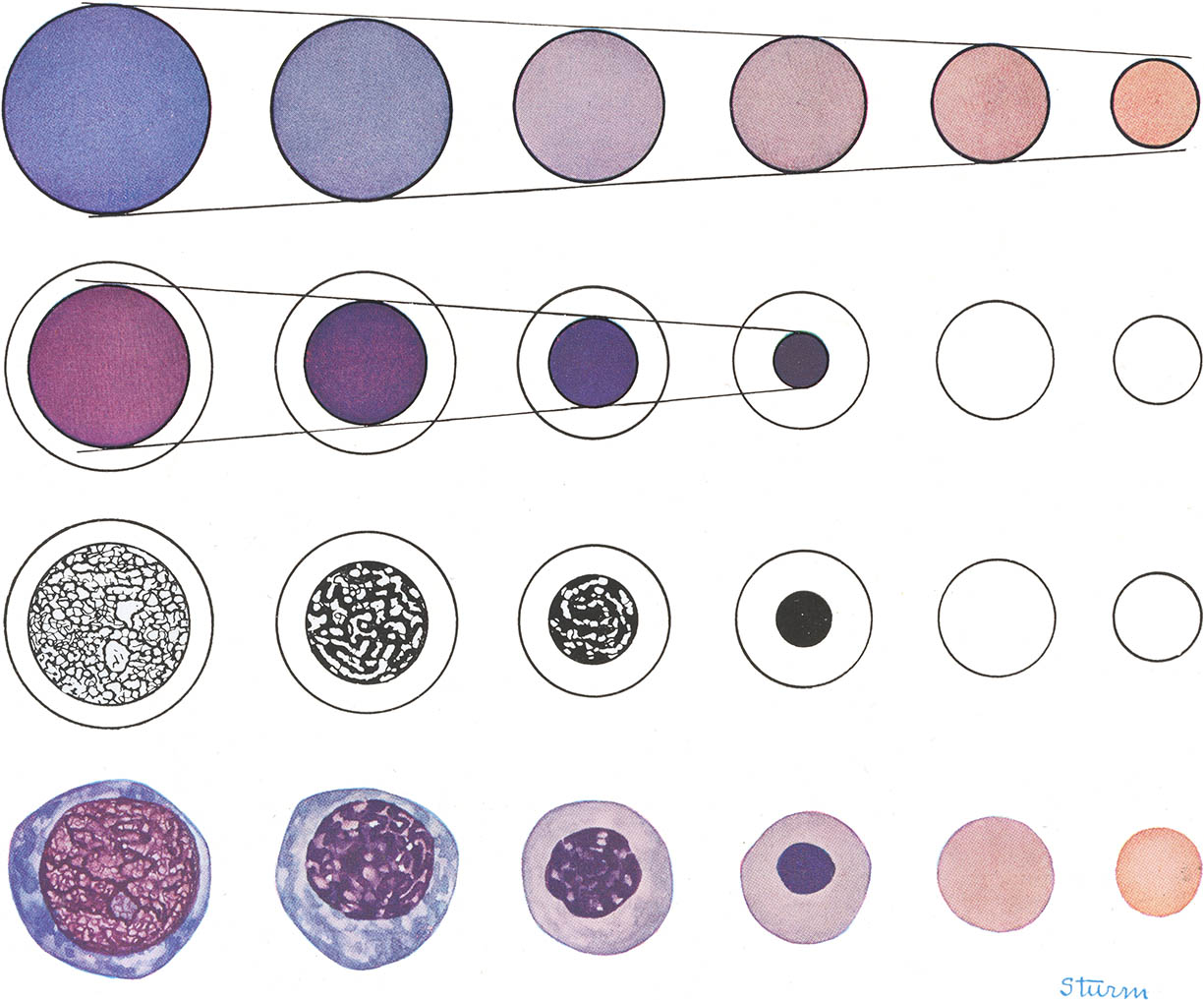
Sturm’s watercolor on paper illustrations, drawn directly from Wright-stained smears prepared by Diggs or Bell, depicted normal, pathological and infectious hematology with a clarity, detail and beauty that microphotography of the 1950s simply couldn’t approach. JAMA, in a review of the first edition, even called her work “of exceptional quality.”
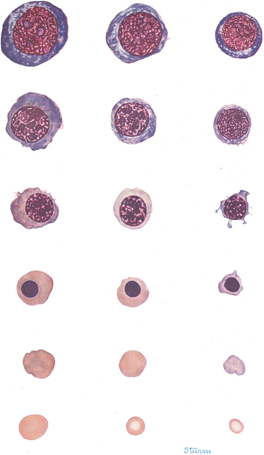
Erythrocytic system
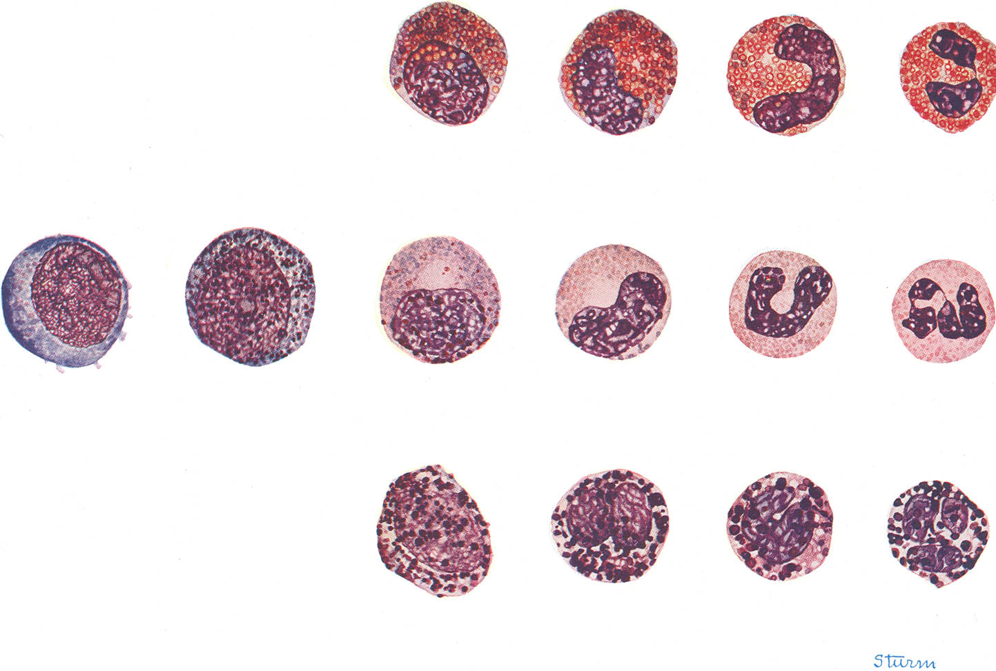
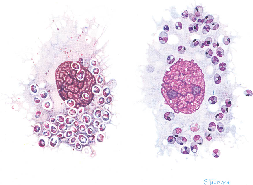
Granulocytic (myelocytic) system
Megakarocytes and thrombocytes
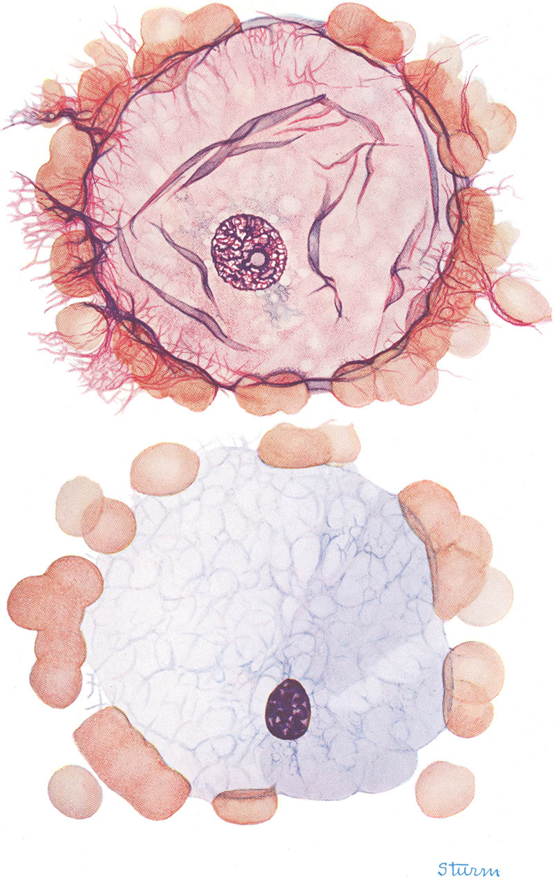
Fat cells
Morphology turned out to be an unlikely classic. It was eventually published by Abbott and went through seven editions and five translations. Sturm’s illustrations survived until the last edition. They would lie beside the microscope in a lab or on the desk of a medical student for nearly 50 years.
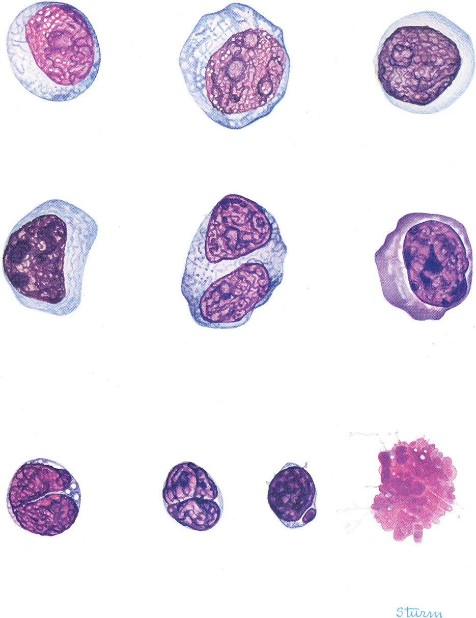
Lymphocytic leukemia
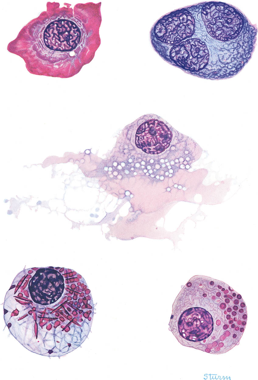
Pathologic plasmocytes
Blood parasites
In her long career Sturm worked in watercolor, tempera, collage, stained glass and sculpture. After Morphology she mostly gave up medical illustration and concentrated on enamels (which made her nationally famous – at least in artistic enamel circles). She died in Memphis in 1988.
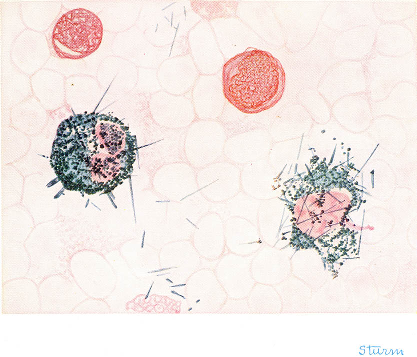
Peroxidase (Sato and Sekiya) stain
1. As a final nod to the armchair hematologist all of these scans (from the first edition, see ref. 4) are identically scaled. So yes, fat cells are really that fat.
2. Memphis, Tennessee. Home of Graceland, but you already know this.
3. “Artist's First Hand Drawings Lend Helping Hand to Surgery.” 23 Jul 1936. The article was picked up by many papers, including the Southeast Missourian.
4. Dr. Lemuel W. Diggs, M.A, M.D., or according to Ann Bells memorial: physician, medical educator, hematologist, clinical investigator, blood banker, researcher, speaker, author, humanitarian, father, grandfather and farmer.
5. Diggs, L. W., Sturm, Dorothy, Bell, Ann. The Morphology of Human Blood Cells. Philadelphia: W. B. Saunders, 1956. The last edition was Bell, Ann, Sallah, Sabah, Diggs, L. W. The Morphology of Human Blood Cells. Abbott Park, Illinois: Abbott Laboratories, 2005. In addition to the seven editions of the book Abbott also published a series of large-format paperback monographs with many of the illustrations. Of all of these possibilities the first edition is the one to get:
True story: when your humble narrator was about 10 and in his “lets be a scientist” phase, one of the Abbott monographs was one of my favorites. Like many things it faded off into the mists of time. Recently, when I vaguely remembered the book (but none of the details), I though it would remain just that – lost to time. Turns out it took all of three minutes on Google to identify the book and purchase the first edition. I am quite serious when I say that this internet thing is going to catch on!
5 Oct 2011 ‧ Anatomicae